What is an Abdominal Aortic Aneurysm (AAA)?
An Abdominal Aortic Aneurysm (AAA) is an aneurysm a bulge or swelling in a blood vessel. Your doctor has determined that a portion of the aorta passing through your abdomen - the area between your legs and your chest - has developed a blood clot. In most cases these clots are caused by fatty deposits that build up inside the arteries.
Aneurysms are dangerous because the blood clot weakens the blood vessel and can cause it to burst. The surgery your doctor has recommended will remove the blood clot and reinforce the weakened wall of the aorta.
This type of bulge occurs when a blood clot or blood clots develop in the aorta, causing to expand. In your case, your doctor has determined that a portion of the aorta passing through your abdomen - the area between your legs and your chest - has developed a blood clot. In most cases these clots are caused by fatty deposits that build up inside the arteries.
Aneurysms are dangerous because the blood clot weakens the blood vessel and can cause it to burst. The surgery your doctor has recommended will remove the blood clot and reinforce the weakened wall of the aorta.
After you are unconscious, your doctor will make a vertical incision down the center of your abdomen. Skin and other tissue will be pulled back in order to expose the abdominal muscles. Your doctor will carefully divide the muscle in order to expose the abdominal cavity.
A special instrument called a retractor will be used to hold the chest open. Once your doctor has a clear view of the abdomen he or she will gently pull the intestines up and out of the way revealing the aorta and the aneurysm.
Now your doctor can begin to remove the clot. First, he or she will apply clamps to each of the two arteries that branch away from the main artery - temporarily preventing blood from flowing to your legs. Next, your doctor will clamp the artery above the aneurysm.
Once the blood supply has been shut off in this manner, your doctor will make a vertical incision in the artery wall and two small horizontal incisions to allow access to the damaged area. The blood clot can then be removed.
The surgical team will sew together any damaged blood vessels inside the aorta. A tube made of a sterile synthetic material can now be inserted into the vessel to provide support and reinforcement. It is then sewn into place.
One by one your doctor will remove the clamps, restoring blood flow to the legs. After verifying there are no leaks around the surgical field, the team will finally close the vessel with sutures. Your doctor will restore all internal organs to their proper positions. The muscles and other tissue can then be closed with sutures. Finally, the skin is closed with staples and a sterile dressing is applied.
Cardiac Catheterization Angiography
There is no radioactive material that will be introduced into the artery of the patient. Contrast Media is not a radioactive material. Contrast Media is a special dye with high atomic number and can be introduced intravenously where it is used to better visualize the vessels because these vessels are in low atomic number. So introduction of high atomic no. contrast media is needed to be able to visualize these vessels.
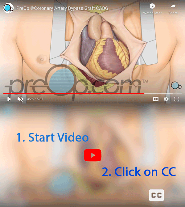
Coronary Artery Bypass Graft (CABG)
magnificent..i don't know what i want to say but anyway i thank the man who did that video and who post it on youtube..#thank_you
Coronary Artery Bypass Graft (CABG off-pump)
I got operated coronary bypass surgery before four years back,now I am quite o.k.


